 Movie
Movie Controller
 Structure viewers
 About EMN search


-Search query
-Search result
Showing 1 - 50 of 62 items for (author: roux & a)
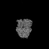
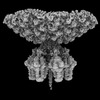
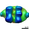
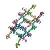
EMDB-17350: 
Single particle cryo-EM co-structure of Klebsiella pneumoniae AcrB with the BDM91288 efflux pump inhibitor at 2.97 Angstrom resolution
Method: single particle / : Boernsen C, Mueller RT, Pos KM, Frangakis AS
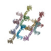
PDB-8p1i:
Single particle cryo-EM co-structure of Klebsiella pneumoniae AcrB with the BDM91288 efflux pump inhibitor at 2.97 Angstrom resolution
Method: single particle / : Boernsen C, Mueller RT, Pos KM, Frangakis AS

EMDB-36794:
Cryo-EM structure of Na+,K+-ATPase alpha2 from Artemia salina in cation-free E2P form
Method: single particle / : Abe K, Artigas P

PDB-8k1l:
Cryo-EM structure of Na+,K+-ATPase alpha2 from Artemia salina in cation-free E2P form
Method: single particle / : Abe K, Artigas P
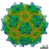
EMDB-41649:
P22 Mature Virion tail - C6 Localized Reconstruction
Method: single particle / : Iglesias S, Cingolani G, Feng-Hou C
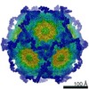
EMDB-41651:
In situ cryo-EM structure of bacteriophage P22 portal protein: head-to-tail protein complex at 3.0A resolution
Method: single particle / : Iglesias SM, Cingolani G, Feng-Hou C
EMDB-41819:
In situ cryo-EM structure of bacteriophage P22 tailspike protein complex at 3.4A resolution
Method: single particle / : Iglesias SM, Feng-Hou C, Cingolani G

PDB-8tvr:
In situ cryo-EM structure of bacteriophage P22 tail hub protein: tailspike protein complex at 2.8A resolution
Method: single particle / : Iglesias S, Cingolani G, Feng-Hou C
PDB-8tvu:
In situ cryo-EM structure of bacteriophage P22 portal protein: head-to-tail protein complex at 3.0A resolution
Method: single particle / : Iglesias SM, Cingolani G, Feng-Hou C
PDB-8u1o:
In situ cryo-EM structure of bacteriophage P22 tailspike protein complex at 3.4A resolution
Method: single particle / : Iglesias SM, Feng-Hou C, Cingolani G
EMDB-41791:
In situ cryo-EM structure of bacteriophage P22 gp1:gp4:gp5:gp10:gp9 N-term complex in conformation 1 at 3.2A resolution
Method: single particle / : Iglesias S, Feng-Hou C, Cingolani G
EMDB-41792:
In situ cryo-EM structure of bacteriophage P22 gp1:gp5:gp4: gp10: gp9 N-term complex in conformation 2 at 3.1A resolution
Method: single particle / : Iglesias S, Feng-Hou C, Cingolani G

PDB-8u10:
In situ cryo-EM structure of bacteriophage P22 gp1:gp4:gp5:gp10:gp9 N-term complex in conformation 1 at 3.2A resolution
Method: single particle / : Iglesias S, Feng-Hou C, Cingolani G

PDB-8u11:
In situ cryo-EM structure of bacteriophage P22 gp1:gp5:gp4: gp10: gp9 N-term complex in conformation 2 at 3.1A resolution
Method: single particle / : Iglesias S, Feng-Hou C, Cingolani G

EMDB-15802:
T5 Receptor Binding Protein pb5 in complex with its E. coli receptor FhuA
Method: single particle / : Degroux S, Effantin G, Linares R, Schoehn G, Breyton C

PDB-8b14:
T5 Receptor Binding Protein pb5 in complex with its E. coli receptor FhuA
Method: single particle / : Degroux S, Effantin G, Linares R, Schoehn G, Breyton C
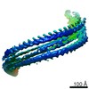
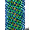
EMDB-10136:
Double-stranded helical ESCRT-III filament formed from Snf7/Vps24/Vps2 on a helical membrane bicelle
Method: helical / : Frost A, Johnson I, Talledge N
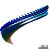
EMDB-10137:
Refined, asymmetrically masked double-stranded helical ESCRT-III filament formed from Snf7/Vps24/Vps2 on helical lipid bicelle
Method: helical / : Frost A, Johnson I, Talledge N
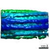
EMDB-10138:
Segment of helical membrane tube with longitudinal ESCRT-III filaments with different binding modes formed from Snf7/Vps24/Vps2
Method: subtomogram averaging / : Moser von Filseck J, Roux A
EMDB-10139:
Segment of helical membrane tube with longitudinal ESCRT-III filaments in the equatorial binding mode formed from Snf7/Vps24/Vps2
Method: subtomogram averaging / : Moser von Filseck J, Roux A
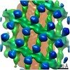
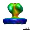
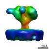
EMDB-4584:
Structure and assembly of the mitochondrial membrane remodelling GTPase Mgm1
Method: subtomogram averaging / : Faelber K, Dietrich L, Noel J, Wollweber F, Pfitzner A, Muehleip A, Sanchez R, Kudryashev M, Chiaruttin N, Lilie H, Schleger J, Rosenbaum E, Hessenberger M, Matthaeus C, Noe F, Roux A, van der Laan M, Kuehlbrandt W, Daumke O
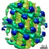
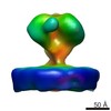
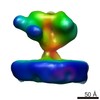
EMDB-10062:
Structure of s-Mgm1 decorating the outer surface of tubulated lipid membranes
Method: subtomogram averaging / : Faelber K, Dietrich L, Noel JK, Sanchez R, Kudryashev M, Kuehlbrandt W, Daumke O
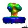
EMDB-10063:
Structure of s-Mgm1 decorating the outer surface of tubulated lipid membranes in the GTPgammaS bound state
Method: subtomogram averaging / : Faelber K, Dietrich L, Noel JK, Sanchez R, Kudryashev M, Kuelbrandt W, Daumke O
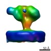
EMDB-10064:
Structure of s-Mgm1 decorating the inner surface of tubulated lipid membranes
Method: subtomogram averaging / : Faelber K, Dietrich L, Noel JK, Sanchez R, Kudryashev M, Kuelbrandt W, Daumke O
EMDB-10065:
Structure of s-Mgm1 decorating the inner surface of tubulated lipid membranes in the GTPgammaS bound state
Method: subtomogram averaging / : Faelber K, Dietrich L, Noel JK, Sanchez R, Kudryashev M, Kuelbrandt W, Daumke O
PDB-6rzt:
Structure of s-Mgm1 decorating the outer surface of tubulated lipid membranes
Method: subtomogram averaging / : Faelber K, Dietrich L, Noel JK, Sanchez R, Kudryashev M, Kuehlbrandt W, Daumke O
PDB-6rzu:
Structure of s-Mgm1 decorating the outer surface of tubulated lipid membranes in the GTPgammaS bound state
Method: subtomogram averaging / : Faelber K, Dietrich L, Noel JK, Sanchez R, Kudryashev M, Kuelbrandt W, Daumke O
PDB-6rzv:
Structure of s-Mgm1 decorating the inner surface of tubulated lipid membranes
Method: subtomogram averaging / : Faelber K, Dietrich L, Noel JK, Sanchez R, Kudryashev M, Kuelbrandt W, Daumke O
PDB-6rzw:
Structure of s-Mgm1 decorating the inner surface of tubulated lipid membranes in the GTPgammaS bound state
Method: subtomogram averaging / : Faelber K, Dietrich L, Noel JK, Sanchez R, Kudryashev M, Kuelbrandt W, Daumke O


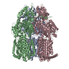
EMDB-0007:
MlaBDEF complex from A. baumannii
Method: single particle / : Bergeron JRC, Kollman JM

PDB-6ic4:
Cryo-EM structure of the A. baumannii MLA complex at 8.7 A resolution
Method: single particle / : Bergeron JR, Kollman JM

EMDB-3718:
3,4-dihydroxybenzoate decarboxylase AroY from Enterobacter cloacae in the apo state
Method: single particle / : Baerland N, Kaltwasser S, Vonck J, Pavkov-Keller T
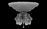
EMDB-6538:
Structure of Simian Immunodeficiency Virus Envelope Spikes bound with CD4 and Monoclonal Antibody 36D5
Method: subtomogram averaging / : Hu G, Liu J, Roux KH, Taylor KA
EMDB-6539:
Structure of Simian Immunodeficiency Virus Envelope Spikes bound with CD4 and Monoclonal Antibody 36D5
Method: subtomogram averaging / : Hu G, Liu J, Roux KH, Taylor KA
EMDB-6540:
Structure of Simian Immunodeficiency Virus Envelope Spikes bound with CD4 and Monoclonal Antibody 36D5
Method: subtomogram averaging / : Hu G, Liu J, Roux KH, Taylor KA
EMDB-6541:
Structure of Simian Immunodeficiency Virus Envelope Spikes bound with CD4 and Monoclonal Antibody 36D5
Method: subtomogram averaging / : Hu G, Liu J, Roux KH, Taylor KA
EMDB-6542:
Structure of Simian Immunodeficiency Virus Envelope Spikes bound with CD4 and Monoclonal Antibody 36D5
Method: subtomogram averaging / : Hu G, Liu J, Roux KH, Taylor KA
EMDB-6543:
Structure of Simian Immunodeficiency Virus Envelope Spikes bound with CD4 and Monoclonal Antibody 36D5
Method: subtomogram averaging / : Hu G, Liu J, Roux KH, Taylor KA
PDB-3jcb:
Structure of Simian Immunodeficiency Virus Envelope Spikes bound with CD4 and Monoclonal Antibody 36D5
Method: electron tomography / : Hu G, Liu J, Roux K, Taylor KA
PDB-3jcc:
Structure of Simian Immunodeficiency Virus Envelope Spikes bound with CD4 and Monoclonal Antibody 36D5
Method: electron tomography / : Hu G, Liu J, Roux K, Taylor KA

EMDB-3112:
Negative stain EM structure of the Tse6-Tsi6-VgrG1-EF-Tu complex in detergent
Method: single particle / : Whitney J, Quentin D, Sawai S, LeRoux M, Harding B, Ledvina H, Tran B, Robinson H, Goo YA, Goodlett D, Raunser S, Mougous J

EMDB-3113:
Negative stain EM structure of the Tse6-Tsi6-VgrG1-EagT6-EF-Tu complex
Method: single particle / : Whitney J, Quentin D, Sawai S, LeRoux M, Harding B, Ledvina H, Tran B, Robinson H, Goo YA, Goodlett D, Raunser S, Mougous J

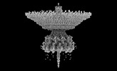
EMDB-1813:
Structural Comparison of HIV-1 Envelope Spikes with and without the V1V2 Loop.
Method: subtomogram averaging / : Hu G, Liu J, Taylor KA, Roux KH

EMDB-1814:
Structural Comparison of HIV-1 Envelope Spikes with and without the V1V2 Loop
Method: subtomogram averaging / : Hu G, Liu J, Taylor KA, Roux KH

EMDB-1519:
Cryoelectron tomography of HIV-1 envelope spikes
Method: electron tomography / : Zhu P, Winkler H, Chertova E, Taylor KA, Roux KH
EMDB-1596:
HIV-1 Env spike maps using various alignment and classfication combinations
Method: subtomogram averaging / : Zhu P, Winkler H, Chertova E, Taylor KA, Roux KH
Pages:
 wwPDB to switch to version 3 of the EMDB data model
